In this blog, we delve into one of the prominent application of Thermal Imaging for detection of breast cancer in a non-invasive, portable, and affordable manner. While the roots of thermal imaging for breast cancer detection trace back to the 1960s, its journey has been marked by fluctuating interest and contradictory findings, primarily due to a lack of standardization and ease of use.
Fast forward to the present decade, and we witness a resurgence of interest in thermal imaging, fueled by the strides made in machine learning. The convergence of technological advancements and the rising incidence of breast cancer has thrust thermal imaging into the spotlight as a potential game-changer. In this blog, we aim to be your guide, providing essential insights and knowledge to prepare you for a deep dive into the world of thermal imaging for early breast cancer detection.
1. Why Breast Cancer?
Breast cancer continues to be the leading cause of cancer-related fatalities among women globally. Remarkably, even in the present day, every alternate woman diagnosed with breast cancer is succumbing to the disease in low and middle-income countries (LMICs). According to the latest data from GLOBOCAN, a staggering 685,000 women lost their lives to breast cancer in 2020. While developed countries have successfully implemented mammography screening programs to combat high mortality rates, LMICs face formidable challenges in adopting these practices. Issues such as exorbitant equipment costs, the demand for specialized expertise, and the necessity for dedicated infrastructure hinder the implementation of conventional breast imaging methods in these regions. Consequently, there is a notable absence of widely employed imaging modalities for breast cancer screening, resulting in delayed detection and treatment.
Adding to the complexity of the situation, the incidence of breast cancer in young women is on the rise worldwide. Unfortunately, there is currently no recommended screening modality specifically tailored for young women, as mammography exhibits low sensitivity in detecting cancers in dense breast tissue. This underscores the urgent need for innovative and accessible screening methods that can address the unique challenges posed by breast cancer in LMICs and cater to the increasing incidence of the disease in younger populations globally.

2. Rise of Thermal Imaging for Breast Cancer Detection
In recent years, thermal imaging has emerged as a promising modality for the early detection of breast cancer. The adoption of thermal imaging in breast cancer detection presents a myriad of advantages, ranging from its cost-effectiveness to its portability and radiation-free characteristics, making it a suitable modality for LMICs. The inherent features such as privacy-aware, non-invasive, no-touch and no-see screening could contribute to increased patient comfort and acceptance, encouraging more individuals to undergo regular screenings. The recent research demonstrated that thermography combined with AI is effective even for young women or women with dense breasts
For a more in-depth exploration of the principles and applications of thermal imaging, refer to my previous blog post titled Introduction to Thermal imaging. This comprehensive resource delves into the foundations of thermal imaging, shedding light on its principles and its potential applications.
3. Biological Relation between Breast Cancer and Thermal Patterns
Cancer is characterized by uncontrolled cellular growth and necessitates increased resource consumption. To facilitate this, cancer cells release Nitric Oxide into the blood, causing alterations in microcirculation. Coupled with the aggressive nature of cancer, this process intensifies blood circulation by dilating vessels, giving rise to new blood vessels (neo-angiogenesis) and the recruitment of dormant vessels.
The substantial large volume blood flow within these vessels connected to the tumor results in elevated temperatures compared to normal blood vessels. This heightened blood flow not only distorts vessel structures but also raises the tumor temperature in comparison to neighboring tissues. Experimental evidence using contact temperature measurements confirms these observations.


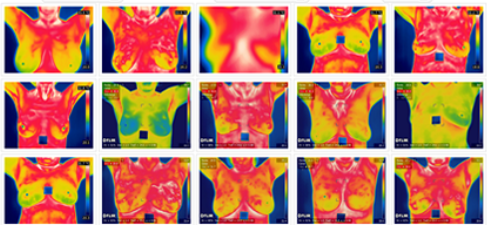
Thermal Image showing a high thermal activity on the left breast in Lower Outer Quadrant (LOQ)
4. Types of Breast Thermography
Static Thermography
Static thermography involves the capture of thermal images at specific view angles after a cooling period, typically around 10 minutes. During the cooling period, the patient's body temperature stabilizes, which allows for dissipation of extraneous heat. Some studies suggest that external cooling even acts as a contrasting agent, constricting vessels connected to the nervous system (sympathetic vessels) and reducing blood supply. Notably, vessels formed due to cancerous growth are not sympathetic, resulting in a temperature disparity between cancerous activity and normal tissue.
After this stabilization, thermal images are acquired from specific angles: Frontal (0°), Left Lateral (+90°), Right Lateral (-90°), Left Oblique (45°), and Right Oblique (-45°). This multi-angle approach ensures complete coverage of the entire breast region. These five images are analyzed for any abnormal focal and vascular changes, facilitating the detection of potential breast malignancies.

Dynamic Thermography
Dynamic breast thermography involves the capture of a series of thermal images or a video over a specific duration to observe and analyze thermal variations over time. Unlike static thermography, which focuses on specific view angles after a cooling period, dynamic thermography provides a continuous recording of temperature changes, offering a more comprehensive understanding of breast physiology.
During dynamic thermography, the patient undergoes a controlled warming phase, often induced by a cooling. As discussed, this process stimulates blood flow, emphasizing the dynamic response of the vascular system. The recorded thermal patterns during this warming phase can reveal information about the vascularity and hemodynamic activity in the breast tissue. This continuous monitoring can aid in distinguishing between normal variations and pathological conditions, contributing to a more nuanced diagnostic approach. In some research studies, it has been discovered that dynamic thermography can effectively differentiate between types of cancers and inflammation. This capability arises because different abnormalities significantly influence the release of Nitric Oxide cycles, resulting in distinct heat patterns.
Though dynamic thermography has advantages, it requires lot more memory and computational power to analyze as even a minute of video could be as high as 1 GB. The interpretation involves assessing temperature changes over time and correlating them with specific physiological processes.
5. Visual Interpretation
Visual interpretation of thermal images involves analyzing the thermal patterns in the captured thermal image to identify for any abnormal focal or vascular changes. Ville-Marie IR Scale is one of the famous breast thermal imaging grading scale that is available out there for visual interpretation of breast thermal images. It uses both vascular and focal temperatures to grade the thermal images into 1-5 grading. A grade of 1 indicates low likelihood of cancerous activities and a grade of 5 indicates high likelihood of cancerous activities.
However, as discussed in our earlier blog- AI over Thermal Images for Medical Applications: Thermal Imaging History and Manual Interpretation Challenges, poses challenges due to the expertise required for accurately detecting vascularity and focal temperatures. Moreover, the manual interpretation process is subjective and demanding for healthcare professionals. The high expertise required for manual interpretation has resulted in conflicting findings regarding the efficacy of thermal imaging for breast cancer detection. Varied research outcomes have presented divergent views, with some studies demonstrating high accuracies while others indicate poor performance.

6. AI Problem Statements
In response to the above challenges with visual interpretation, there has been a notable surge in research over the last decade, focusing on the integration of artificial intelligence (AI) to streamline and enhance the visual interpretation of thermal images in medical applications. AI has the potential to mitigate the subjectivity and complexity associated with manual interpretation, offering a more efficient and accurate method for detecting abnormal thermal patterns indicative of potential health issues. In addition, in the last five years, a multitude of start-ups have emerged, actively conducting clinical evaluations to assess the feasibility of employing AI in conjunction with breast thermography.
Ongoing research endeavors are focusing on various areas to position breast thermography as a frontline modality for detecting breast cancer. Let's delve into the existing AI problem statements in this novel field of malignancy detection from thermal images.
Classification of Breast Cancer from Thermal Images:
- Objective: Classifying breast thermal images, whether static or dynamic, into normal, benign, or malignant categories.
- Significance: Enhancing accuracy in categorizing thermal images aids in early identification and intervention.
Segmentation of Breast Region of Interest:
- Objective: Accurately isolating the breast region from thermal images that may capture surrounding areas such as the neck and abdomen.
- Significance: Focused analysis on the breast region is crucial for precise detection and classification of abnormalities.
Localization of Abnormal Regions in Thermal Images:
- Objective: Identifying abnormal regions, such as hotspots and vascular structures, within thermal images.
- Significance: Facilitating targeted examinations, helping doctors plan interventions for a comprehensive understanding of detected irregularities.
3D Surface Generation:
- Objective: Addressing the challenge of correlating heat patterns across multiple views by generating 3D breast surface from discrete 3D views.
- Significance: Provides a more comprehensive visualization of breast thermography data for enhanced diagnostic capabilities.
3D Internal Modeling:
- Objective: Simulating internal heat patterns within the breast tissue from captured 2D thermal images.
- Significance: Overcoming the limitation of surface-level information in thermal imaging, aiding in the study of internal anatomical changes crucial for detecting cancerous lesions.
These problem statements represent critical research directions aimed at advancing the capabilities of AI in conjunction with breast thermography, paving the way for more effective and reliable breast cancer detection methods.
7. Open Source Datasets
Below are two open source datasets which allows you develop algorithms for the above AI problem statements.
Database For Mastology Research (DMR) for Breast Cancer Detection
DMR database is an open source dataset from Visual Lab, Universidade Federal Fluminense. It consists of breast thermal imaging data of 200+ participants along with their medical records and diagnosis from standard imaging tests such as mammography. Each participant data include breast thermal images at five standard views along with series of thermal images capture over different time stamps during the imaging.
Dataset Link: http://visual.ic.uff.br/dmi/

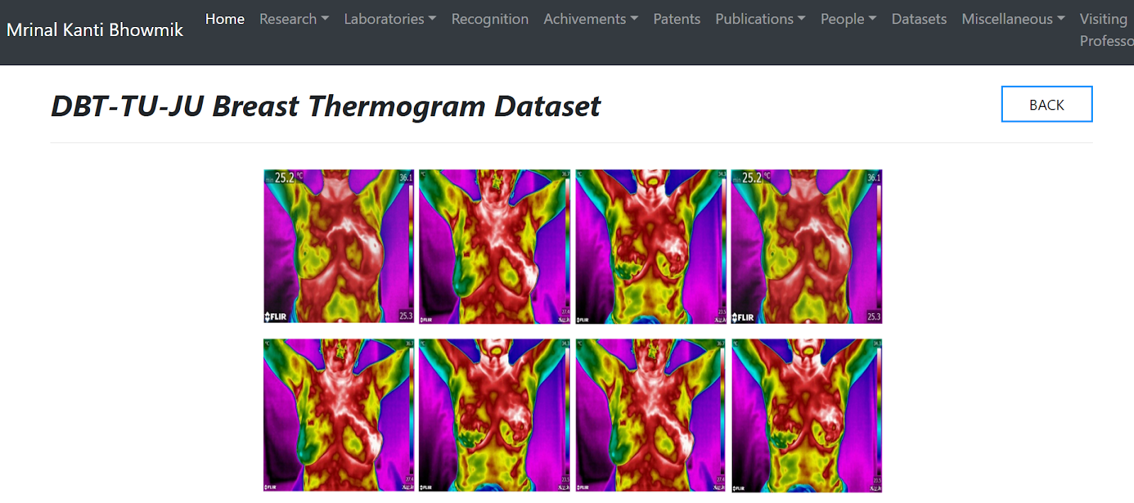
DBT-TU-JU Breast Thermogram Dataset
DBT-TU-JU Breast Thermogram dataset is from Regional Cancer Centre (RCC), Agartala Government Medical College (AGMC) of Govind Ballav Pant (GBP) Hospital, Agartala, Tripura, India. The includes 1100 thermograms from 100 subjects captured at six different views involving the five standard views and supine view. Clinical findings, X-ray mammography and Fine Needle Aspiration Cytology (FNAC) reports are available to obtain the final clinical diagnosis.
Dataset Link: https://www.mkbhowmik.in/dbtTu.aspx
Important Note: In accordance with the regulations of the US Food and Drug Administration (FDA), breast thermography holds approval solely as an adjunct modality for breast screening and is not sanctioned for standalone screening purposes. Nevertheless, recent research findings have highlighted the potential benefits of integrating machine analysis with thermal imaging to derive more objective and data-driven decisions. It is my sincere hope that forthcoming research endeavors will continue to yield substantial evidence, demonstrating the efficacy of this affordable imaging approach. Such validation could play a pivotal role in reducing mortality rates, particularly in low and middle-income countries (LMICs). The exploration of synergies between machine analysis and thermal imaging holds promise for advancing the field and enhancing the utility of breast thermography as a valuable tool in comprehensive breast health assessments.
Do post your questions in the comments section!
Follow the blog to know more about health-tech applications.
Can you share full link for Ville-Marie IR Scale
ReplyDeleteYou can find the description of Ville-Marie grading at https://ieeexplore.ieee.org/document/844378
DeleteCan thermal imaging detect small tumors?
ReplyDeleteYes. Studies show that it can tumors that are as small as 4-5mm
Delete