Why Thermal Imaging Is Essential for Advancing Healthcare?
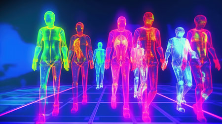
In many developing and underdeveloped countries, providing healthcare services to rural areas still remains a formidable challenge. At the heart of this challenge lies the need for screening technologies that are not only cost-effective but also non-invasive, portable, and require minimal human expertise. Regrettably, many conventional imaging modalities come with exorbitant costs, radiation risks, and a demand for specialized skills. This makes the combination of Artificial Intelligence (AI) and thermal imaging a game changer.
Thermal imaging presents a multitude of benefits: it does not involve any external radiation, offers a no-touch, non-contact experience, boasts portability, and remains remarkably cost-effective. During the COVID-19 pandemic, we all have seen their use extensively in airports, malls, railway stations and other public locations for COVID-19 screening. Apart from this, its usage has been studied for breast cancer screening, diabetic foot, muscular skeletal disorders, pain management, thyroid cancer, oral cancer, skin cancer etc. Its combination with AI also holds the promise of reducing the dependence on scarce human expertise, a critical factor in countries like India, where the ratio stands at just one radiologist for every 100,000 people.
In the inaugural series of our blogs, we embark on a journey to unravel the fundamental concepts of thermal imaging. These insights serve as stepping stones for exploring the transformative potential of AI in the realm of medical applications using thermal imaging.
What is a Thermal Image?
Long wave infrared radiation (LWIR) is naturally emitted by every object, including the human body. Infrared thermal cameras convert this LWIR emitted from an object (or a body) and convert them into a temperature matrix. Each element in the temperature matrix represents a temperature value corresponding to a particular point on the object's surface.

The size and area of the object that is being imaged is determined by the camera's spatial resolution, Field of View (FOV) and its positioning from the object. While the spatial resolution of a thermal camera influences the level of detail captured in the temperature matrix, FOV and camera position decides the area of object. Higher spatial resolution allows for finer granularity and more precise temperature measurements, while lower resolution may result in a coarser representation of the temperature distribution.
The latest advanced thermal cameras have spatial resolution of 1024 × 768 pixels or more with a temperature sensitivity and accuracy error of 20 mK (0.02°C) and ± 1°C . This means that we can measure minute relative temperature differences of up to 0.02°C with a maximum deviation of ± 1°C.
To reach to this level of sophistication, thermal cameras have advanced over the years. If you are interested to know more about the advances in thermal cameras over years, you can read the paper - Kakileti ST, Manjunath G, Madhu H, Ramprakash HV. Advances in breast thermography. New Perspectives in Breast Imaging. Malik A (ed): IntechOpen, London. 2017 Oct 4:91-103.

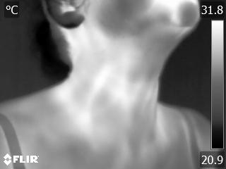
In the context of a human body, the obtained temperature values are real numbers and typically lie between 20℃ and 40℃. Given the continuous range of temperature values, directly studying and analyzing the numerical data with the naked eye becomes impractical. To facilitate visual interpretation and analysis, a mapping function is employed to convert the temperature values into color images a.k.a thermal images that are easier to comprehend. These mapping functions are achieved through the use of color palettes that typically present on the infrared thermal image.
Color Palettes
Color palettes are a predefined sequence of colors that are stacked vertically, forming a visually appealing gradient. The purpose of these color palettes is to establish a correspondence between temperature values and colors, enabling the visualization of temperature variations across the object's surface. To generate a color thermal image from the temperature matrix, a minimum temperature (Tmin) and a maximum temperature (Tmax) are selected as the limits for the color mapping. The temperature range between Tmin and Tmax is divided into multiple bins or intervals, with the number of bins corresponding to the number of colors present in the chosen color palette. Each temperature bin is then mapped to a specific color value in the color palette, resulting in a color image that represents the temperature distribution across the object. Some prominently used color palettes are described below.

Grayscale Palette
Grayscale palette converts the temperature matrix into a gray scale image by mapping the temperature values into a numerical range of 0 to 255, which represents the intensity or brightness of the gray color. Variation in the shades of gray allows one to study the variations in the heat patterns. Cooler temperature regions appear darker or closer to black, while warmer temperature regions appear lighter or closer to white. This grayscale representation allows for a straightforward visualization of temperature differences without the distraction of color information. Grayscale palettes are commonly used for visualizing structural details, such as vessels, bones or tissue boundaries.
Inverted Grayscale Palette
Inverted grayscale palette is an inverse of gray palette formed by stacking the grayscale palette in reverse order. Unlike in the grayscale palette, cooler temperature regions appear lighter or closer to white and warmer temperature regions appear darker or closer to black in inverted grayscale palette. Inverted grayscale images are often of interest in identifying areas of inflammation, blood flow irregularities, and detecting certain medical conditions.

Iron Hot Palette
Iron Hot color palette is a commonly used color palette for visualizing thermal images for industrial applications. However, its usage in medical imaging is very limited. This distinctive palette is characterized by an array of resplendent yellow hues, with the zenith of heat represented by the pristine purity of white.

Camera Parameters for Accurate Temperature Values

Let us see a demo of how these parameters impact the temperature values
Comments
Post a Comment