In this blog, we will delve into the innovative use of thermal imaging for detection of Thyroid Cancer, a cancer which is on rise in the recent years but have excellent prognosis if detected early!!
What is Thyroid Cancer?
Thyroid gland produces hormones that play a crucial role in regulating various bodily functions such as metabolism, heart rate, blood pressure, and body temperature. It is shaped like a butterfly and is situated in the front part of the neck, below the thyroid cartilage, which is also know as Adam's apple. Despite its relatively small size and location, the thyroid gland can develop cancerous cells, leading to the formation of tumors. Thyroid cancer occurs when there is uncontrolled cellular division within the thyroid gland.
The incidence of Thyroid cancers are increasing over the recent years and the incidence rates significantly vary with geography and also gender. Women have 3x more risk of getting Thyroid cancer than men. Identifying thyroid cancer at an early stage is crucial for effective treatment and management. If diagnosed at the early stage, thyroid cancer is one of most curable cancers with overall 5-year survival rate> 99%. This underscores the importance of timely screening and diagnostic measures in identifying thyroid cancer at its nascent stage, thereby improving the prognosis and outcomes for affected individuals.
Thyroid Nodules And Thyroid Cancer
Thyroid nodules are abnormal growths or lumps that develop within the thyroid gland. Having these nodules is common. In most cases, thyroid nodules do not cause any symptoms and are often discovered incidentally during routine physical examinations or imaging studies. Their occurrence is more in women compared to men and increases with age.
However, do not worry!! Most of these thyroid nodules are benign. Only a small fraction thyroid nodules can become cancerous. As per American Cancer Society, only 2 or 3 in 20 nodules are cancerous. However, it is essential to have any new or unusual lumps in the thyroid gland evaluated by a healthcare professional to rule out the possibility of thyroid cancer. The benign nodules may lead to overproduction of thyroid hormones, a condition known as hyperthyroidism, which can cause symptoms such as weight loss, rapid heartbeat, and tremors.
Thermal Imaging for Thyroid Cancer
The uncontrolled cellular growth, a characteristic of cancerous tumors, is often associated with increased blood flow through a process known as angiogenesis. This heightened metabolic activity at the site of the tumor results in the generation of more heat at the vicinity of cancerous cells. As the proximity of the thyroid gland is close to the skin surface, thermal imaging has potential to analyze the metabolic activity associated with the Thyroid gland.
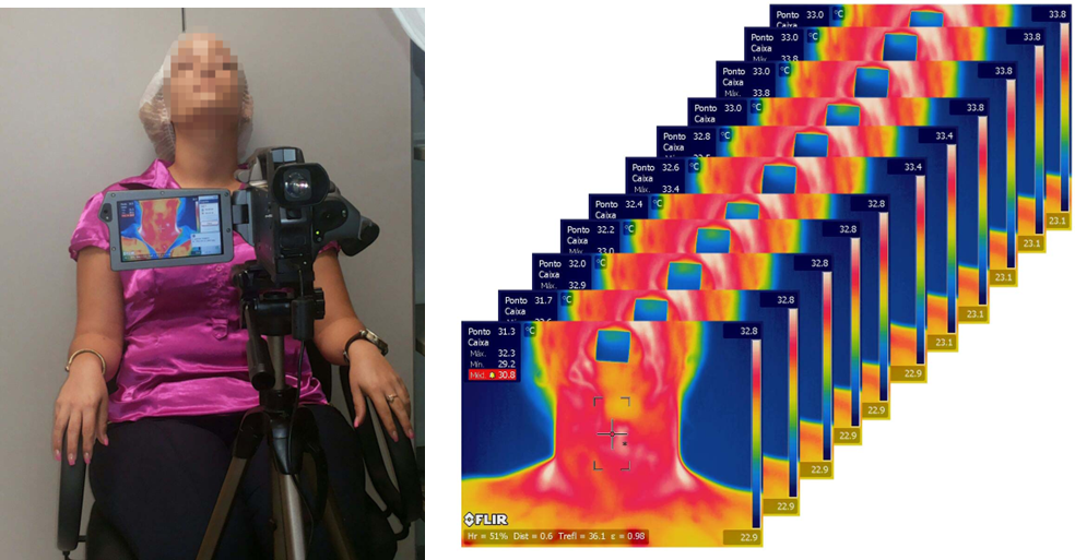
With the advancements in thermal camera technology, modern devices are capable of detecting minute temperature differences as low as 0.05 °C. This level of sensitivity might allow for the visualization and analysis of vascular activity and abnormal thermal patterns associated with cancerous metabolism within the thyroid gland. The below figure shows the thermal activity of cancerous thyroid nodule during warming.

However, there is only a limited research on the application of thermal imaging for thyroid cancer detection. One potential reason for this could be the challenges associated with manual interpretation of thermal images.
Thermal AI for Thyroid Cancer Detection
With the advances in both thermal imaging cameras and artificial intelligence algorithms, multiple research groups are now attempting to develop automated algorithms for detection of Thyroid cancer using the low-cost, portable, non-invasive and radiation-free thermal imaging. Broadly, the AI/ML problems are associated with
Classification of Thyroid Cancer: This involves developing algorithms capable of distinguishing between thermal images associated with thyroid cancer and those that are not. These algorithms may analyze either time-series thermal images or static thermal images to identify patterns indicative of cancerous activity.
Localization of Thyroid Nodules: Another challenge is to accurately locate thyroid nodules within thermal images. By pinpointing the exact location of nodules, clinicians can perform targeted ultrasound examinations or ultrasound-guided fine-needle aspiration cytology (FNAC) to confirm malignancy.
Registration of Time-Series Thyroid Thermal Images: Thermal imaging may capture dynamic changes in temperature over time. Registering or aligning these time-series data points is crucial for tracking changes in thermal patterns associated with thyroid nodules or cancerous growths accurately. Traditional registration algorithms used in RGB space may not be directly applicable to thermal images. Unlike pixel intensity in RGB images, which may remain relatively stable over time, thermal signatures can undergo rapid fluctuations over time due to the continuous metabolic process. As a result, conventional registration techniques do not work and new techniques are required to effectively align thermal data points in time-series images.
Open Source Thermal AI Datasets for Thyroid
The data you post here is not available on the site. Only static images no dynamic once.
ReplyDeleteHi, Dynamic/time series images are also part of the dataset. Incase you are unable to download from their site, please contact the Visual Lab directly. They might share google drive link with all the data.
ReplyDelete